buscar em luizmeira.com

procurar em luizmeira.com

12 dezembro 2010

Re: Mensagem enviada a partir do seu perfil do Google: Iris

Olá Diego,

Estou ao dispor, 

Uma análise rápida, sem abordar exames laboratoriais custa $75,00
luizmeira.com/serve.htm
19  9612 6029


Em 11 de dezembro de 2010 23:14, Diego Marczuk <kyrton.kir@gmail.com> escreveu:
Ola Luiz,
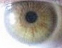
 Vejo seu site e fiquei muito interessado sobre Iridologia, tenho feito estudos sobre desintoxicaçao. Tenho tambem tirado fotos de minha iris durante uma mudança de alimentaçao para notar as mudanças, se puder, gostaria de que analiza-se uma foto de minha ris, grato .

10 dezembro 2010

Sinais

Obrigado pelo estímulo Paulo,

Saudações fraternas
luizmeira.com
19  9612 6029


Em 10 de dezembro de 2010 11:17, Paulo Ricardo Cunha <farmaclara@netsite.com.br> escreveu:
 
FANTÁSTICA,FABULOSA
 -
Date/Time: 2010-12-10 05:17:52 PST
Sender IP: 189.41.187.175 [Brazil] | mm55pai98ulrbo49
Referrer: http://luizmeira.com/irisinais1.htm

27 outubro 2010

Resumo de iridologia

Holla Maya,

Gracias por acordarme deste contenido, me gustaria acerlo mas completo ponendole imagenes.

luizmeira.com
19  9612 6029


2010/10/27 Maya de la Fuente <yuma9@hotmail.com>
 
Excelente.
 --
Date/Time: 2010-10-26 20:26:58 PDT
Sender IP: 187.137.51.199 [Mexico] | mm55pai98ulrbo49
Referrer: http://luizmeira.com/marcos.htm

26 outubro 2010

comentário

Olá Camila,

Obrigado pelo comentário.

luizmeira.com
19  9612 6029


Em 26 de outubro de 2010 19:17, Camila <camillatamarozi@hotmail.com> escreveu:
 
Muitas veses agente sente o nosso corpo e nao sabe o que que é que ajente esa mechendo...precisamos conhecer nosso corpo e por isso nos conhecemos os nossos olhos.
 
 
Date/Time: 2010-10-26 14:17:43 PDT
Sender IP: 189.68.70.222 [Brazil] | mm55pai98ulrbo49
Referrer: http://luizmeira.com/anatomia.htm

02 setembro 2010

análise da íris

enviando as fotos, veja:

luizmeira.com/serve.htm
19  9612 6029

TIM19  8172  0949


Em 1 de setembro de 2010 22:30, Paulo Rodrigues <paulorobmarq@uol.com.br> escreveu:
 
 Fiz uma pesquisa de doenças na internet e achei esse site, estou problemas de saude e gostaria de saber como posso fazer meu diagnóstico através da iris
--
Date/Time: 2010-09-01 18:30:14 PDT
Sender IP: 187.3.33.77 [Brazil] | mm55pai98ulrbo49
Referrer: http://luizmeira.com/irisinais.htm

17 agosto 2010

didática

De: vera <vera.maria45@hotmail.com>
Data: 10 de junho de 2010 16:46

 matéria muito boa...super didático
 --
Date/Time: 2010-06-10 12:46:40 PDT
Sender IP: 187.27.120.232 [Brazil] | mm55pai98ulrbo49
Referrer: http://luizmeira.com/irisinais.htm

21 julho 2010

excelente!

De: cleia dias cruz <cleiadias94@yahoo.com.br>
Data: 21 de julho de 2010 20:39

exelente!

Fiz um curso sobre iridologia em Floripa e agora estou tentando aprender o máximo ,cada informação é muiiito importante para mim, obrigada.


--
Date/Time: 2010-07-21 16:39:51 PDT
Sender IP: 189.74.201.99 [Brazil] | mm55pai98ulrbo49
Referrer: http://luizmeira.com/irisinais.htm

20 julho 2010

análise

Sim Rosângela,

Estou a postos:
luizmeira.com/serve.htm
19  9612 6029
24  9941 3838


Em 20 de julho de 2010 11:48, Rosangela <rosangela1oliveira@yahoo.com.br> escreveu:
 
Gostaria de saber se posso enviar fotos dos meus olhos para o Sr. fazer um avaliação.


--
Date/Time: 2010-07-20 07:47:58 PDT
Sender IP: 189.90.241.22 [Brazil] | mm55pai98ulrbo49
Referrer: http://luizmeira.com/iris.htm

12 julho 2010

nódulo mama

Em 11 de julho de 2010 15:05, @terra.com.br Susan Schneider <susan.schneider> escreveu:

 
A primeira imagem, com nódulo de mama: é a lacuna e não a psora indicada pela seta??
Date/Time: 2010-07-11 11:05:52 PDT
Sender IP: 189.98.170.219 [Brazil] | mm55pai98ulrbo49
Referrer: http://luizmeira.com/iristgu.htm

Olá Susan,

atribuo o nódulo à psora

Atenciosamente
Luiz Meira

08 julho 2010

Loko

Obrigado pelo incentivo Olavo,

Grande Abraço
Luiz Meira


De: Olavo - São Joaquim da Barra SP <olavo_loko@hotmail.com>
Data: 8 de julho de 2010 01:13

 minha mãe estava com uns problemas ai, acho que era hepatite medicamentosa e um medico desse negócio ai falo tudu o que ela tinhe sem ele numca te visto ela e muito menos o estado clínico dela, não conhecia esse estudo mas apartir desse dia fiquei muito impressionado...ele passo uns lance de injeção de sangue dela mesma e indicou os melhores alimentos pra ele conforme seu tipo sanguineo achei animall...
Òtimo site, muito esclarecedor...

Obrigado!


Date/Time: 2010-07-07 21:13:09 PDT
Sender IP: 201.16.216.217 [Brazil] | mm55pai98ulrbo49
Referrer: http://luizmeira.com/iris.htm

21 junho 2010

Proveito


De: franco <franco-sm@hotmail.com>

de muita utilidade agradecido pela gentileza aproveitei bastante.


--
Date/Time: 2010-05-22 10:05:34 PDT
Sender IP: 189.58.211.177 [Brazil] | mm55pai98ulrbo49
Referrer: http://luizmeira.com/irisbody.htm

18 junho 2010

Avaliar a própria íris

Em 18/06/10, DILCE DOMNGUES PINHEIRO
<dilcedp2010/2hotmail.com@mercury.freedback.com> escreveu:

> POSSO EU MESMO ATRAVES DE UMA LENTE DE
> AUMENTO OBSERVAR O ÍRIS, CONSERVANDO EM FOTOS O SEU DESENVOLVIMENTO EM CASOS DE MANCHAS?
>
> Date/Time: 2010-06-18 11:19:10 PDT
> Sender IP: 200.178.193.2 [Brazil] | mm55pai98ulrbo49
> Referrer: http://luizmeira.com/botsaris.htm

Sim Dilce,
Esta é uma das grandes vantagens da iridologia: podemos acompanhar a
evolução dos comprometimentos orgânicos com fotos seriadas da íris ou
mesmo olhando no espelho.

luizmeira.com
19 9612 6029

congratulações

mara

mrkleinubing859@gmail.com

parabens pelo teu site as matérias são ótimas pois sou estudante de
iridologia .e aperfeiçoo meus estudos no teu site .obrigado

Date/Time: 2010-06-18 10:11:07 PDT
Sender IP: 189.126.53.14 [Brazil] | mm55pai98ulrbo49
Referrer: http://luizmeira.com/irisdigest.htm

17 junho 2010

congratulações

coraly n. silviá

carolcromo@hotmail.com

muito muito bom este site, amei.Pretendo em breve me tornar uma
iridóloga muito competente.

Date/Time: 2010-06-17 15:42:41 PDT
Sender IP: 187.58.40.249 [] | mm55pai98ulrbo49
Referrer: http://luizmeira.com/irisdigest.htm

13 junho 2010

didático

vera

vera.maria45@hotmail.com

matéria muito boa...super didático

Date/Time: 2010-06-10 12:46:40 PDT
Sender IP: 187.27.120.232 [Brazil] | mm55pai98ulrbo49
Referrer: http://luizmeira.com/irisinais.htm

29 março 2010

Aparência de Cisto Poplíteo em joelho Direito


iris do dia 18 de julho de 2008


íris em 7 de dezembro de 2008


Popliteal Cyst


Gadgets powered by Google
     Páscoa 2010 Also known as a ‘Popliteal Cyst’, Baker cyst  is a distended bursa caused by knee joint fluid protruding to the back of the knee. It is thus a benign swelling & is named after Dr William Morrant Baker who first described this health condition. The term is a misnomer as it is not a true cyst but is due to synovial fluid distending the bursa. Aetiology- (1) Idiopathic- Baker cysts may sometimes develop without any apparent cause particularly in children. (2) Infection- Local infection may cause a retention of fluid with the subsequent formation of a Baker cyst. (3) Trauma or injury to the knee- It may cause an effusion, thus triggering the formation of a Baker cyst. (4) Arthritis-Arthritis is the most common & osteoarthritis probably the most frequent among arthritides. (5) Internal derangement of knee- Internal derangement of knee like meniscal tears etc. may cause an effusion resulting in the formation of a Baker cyst. Location- It is located posterior to the medial femoral condyle, between the tendons of the medial head of the gastrocnemius & the semimembranosus muscles. Age- Baker cysts appear much less frequently in children than in adults. Pathology- Being an extension of the knee joint, a Baker cyst is a synovial cyst lined with a true synovium. In most cases herniation of synovial membrane through posterior part of capsule takes place. . Escape of fluid through the normal communication of bursa with knee is the other mode. The knee joint effusion caused by intrinsic intra-articular disorders or any other cause is displaced into the popliteal bursa, thus reducing potentially destructive pressure in the joint space. So a Baker cyst may have a protective role to play for the knee. In such cases, the popliteal bursa becomes filled up with fluid & consequently expands resulting in the formation of a swelling. The cyst usually communicates with the joint by eswqweqerqsdaq1y of a slit-like opening or may pinch off. Associated health conditions- Medical conditions associated with Baker cysts are as follows- (1) Arthritis is the most common among which osteoarthritis is the most important. Rheumatoid arthritis, Juvenile rheumatoid arthritis etc are also common. (2) Internal derangement of knee like meniscal tears etc. (3) Infection like septic arthritis. (4) Miscellaneous- Hypothyroidism, Gout, Psoriasis, Systemic lupus erythematosus, Sarcoidosis, Haemophilia, etc. Clinical features- May be asymptomatic or may have the following features in addition to the features of the underlying primary cause- (1) A slight swelling behind the knee which is particularly noticeable on standing & when compared to the opposite uninvolved knee. (2) The swelling is usually soft & fluctuant & is with or without pain. Typically these cysts are not painful unless swelling is extensive. (3) A sensation of tightness behind the knee, especially when the knee is extended or fully flexed. (4) Restricted mobility of the knee joint. (5) Transillumination- Transillumination by a shining light through the cyst may show a mass filled with fluid. (6) In case there is rupture of the cyst, calf tenderness & bruising at the ankle may be present. Investigations- (1) X-ray- An X-ray of the knee joint will not show any cyst, but it may show the presence of other abnormalities which may cause development of a Baker cyst. (2) MRI- An MRI helps to show a cyst with its size & location. (3) Ultrasound- An ultrasound can also determine the location & contents of a cyst. (4) Arthrogram- Arthrograpgy may also be utilized for its detection & it is more sensitive than ultrasonography in its detection. Complications- A Baker cyst may sometimes compress vascular structures & may cause a deep vein thrombosis. It may also rupture & cause extravasation of fluid in the calf. There may also be haemorrhage into the cyst in some cases, particularly if there is any associated bleeding disorder. Infection in case of a Baker cyst is very rare. Differential Diagnosis- A Baker cyst may sometimes be confused with thrombophlebitis or deep vein thrombosis from which it is to be differentiated by urgent blood tests & other investigations. It may also sometimes be confused with septic arthritis or a ganglion cyst. Treatment- [A] General measures to be taken are- (1) Treatment of underlying cause like arthritis or torn knee cartilage. (2) Temporarily avoiding activities that may increase the load on the knee joint. (3) Physiotherapy. (4) Exercises to maintain mobility & strength of the knee joint. [B] Homeopathic medicines to be used – Homeopathy can be very effective if properly used. Homeopathic medicines to be used depend on the size of the cyst along with its cause & the symptoms produced. Ruta Graveolens (Ruta), Rhus toxicodendron (Rhus tox), Bryonia etc. may be used. The potency & frequency of dosage as well as duration of treatment varies with the severity of the condition & the individual Prevention- Prevention of knee injury is essential for reducing the risk of development of a Baker cyst for the first time or its recurrence after treatment. Hence supportive footwear appropriate to the activity of an individual is to be worn as well as stoppage of the activity & seeking of medical advice after an injury is needed. Prognosis- Prognosis of Baker cysts depends on the presence of any underlying knee pathology & the degree of its response to treatment? Most Baker cysts without any underlying knee pathology disappear spontaneously after several years, particularly in children & young adults in whom usually there is no underlying knee pathology? But in some cases a Baker cyst continues to grow with worsening of the symptom & ultimately may rupture & produce acute pain behind the knee & in the calf & swelling of the calf muscles? In short, a Baker cyst manifests itself as a soft swelling behind the knee with or without pain & can be treated by Ruta, Rhus tox or Bryonia. But in every case, a doctor should be consulted.